Acromioclavicular Joint Injury, A Simple Guide To The Condition, Diagnosis, Treatment And Related Conditions
Par :Formats :
Disponible dans votre compte client Decitre ou Furet du Nord dès validation de votre commande. Le format ePub est :
- Compatible avec une lecture sur My Vivlio (smartphone, tablette, ordinateur)
- Compatible avec une lecture sur liseuses Vivlio
- Pour les liseuses autres que Vivlio, vous devez utiliser le logiciel Adobe Digital Edition. Non compatible avec la lecture sur les liseuses Kindle, Remarkable et Sony
Notre partenaire de plateforme de lecture numérique où vous retrouverez l'ensemble de vos ebooks gratuitement
Pour en savoir plus sur nos ebooks, consultez notre aide en ligne ici
C'est si simple ! Lisez votre ebook avec l'app Vivlio sur votre tablette, mobile ou ordinateur :

- FormatePub
- ISBN978-0-463-98078-1
- EAN9780463980781
- Date de parution08/01/2020
- Protection num.pas de protection
- Infos supplémentairesepub
- ÉditeurBluewater
Résumé
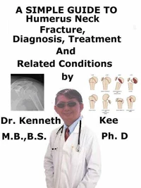
This book describes Acromioclavicular Joint Injury, Diagnosis and Treatment and Related DiseasesThe acromioclavicular (AC) joint injury is an injury to the acromioclavicular (AC) joint with disruption of the AC ligaments with or without coracoclavicular (CC) ligament disruption. Acromioclavicular Joint Injury is a frequent injury among athletes and young persons. AC injuries are often seen after sporting events, car accidents, falls from a bicycle, and other sports-related activities (e.g., skiing).
AC joint injuries are responsible for more than 40% of all shoulder injuries and nearly 10% of all injuries in collision sports such as football, lacrosse, and ice hockeyAcromioclavicular injuries may be linked with a fractured clavicle, impingement syndromes, and more rarely neurovascular injuries. The most frequent mechanism of injury is direct trauma to the lateral shoulder or acromion process with the arm in adduction.
Falling on an outstretched hand or elbow may also cause AC joint separation. A direct blow to the shoulder may cause AC joint injury, often sustained while falling onto the shoulderThere is pain normally over AC joint which can also be extended to the trapeziusPatients with an AC joint injury normally manifest with shoulder pain, normally superior-anterior in location, and will depict a mechanism of injury indicating this type of injury.
They may depict pain spreading to the neck or shoulder which is often worse with movement or when they attempt to sleep on the affected shoulder. On physical examination, the doctor may notice swelling, bruising, or a deformity of the AC joint depending on the degree of injury. The patient will feel tenderness at that location. The doctor can evaluate the stability of the AC joint with anterior-posterior mobility (acromioclavicular ligament) and vertical mobility (coracoclavicular ligaments).
Standard x-rays are sufficient to make a diagnosis of acromioclavicular joint injury and should be used to assess for other causes of traumatic shoulder pain. AC joint injuries may not always be evident on normal radiographic views (anteroposterior AP, lateral). Additional X-rays views are:1. The zanca view, an AP view done by tilting the beam 10 to 15 degrees cranial, and2. Bilateral AP views to compare displacement to the contra-lateral shoulder.
Weighted stress views may be done to assess the displacement of the joint when the diagnosis is not certain on standard AP views. It is important to appraise the entire clavicle for possible fracture or sterno-clavicular injury as well as do a full neurovascular examination on the affected extremity. Non-operative treatment comprises brief sling, immobilization, rest, ice, physical therapyIndications for non-operative treatment are:1.
Type I and II2. Type III in most personsGood results are obtained when the clavicle is displaced < 2cmSurgical treatment is needed for:1. Acute type IV, V or VI injuriesRecent studies indicate no disparity in functional outcomes between operative and non-operative interventions for high grade injuries2. Acute type III injuries in laborers, elite athletes, patients with cosmetic worries3. Chronic type III injuries that failed non-operative treatmentAcute injuries were treated with ORIF (open reduction internal fixation) and chronic injuries were treated with CC ligament reconstructionNew studies have shown no disparity in outcomes in types III injuries treated surgically after 6 weeks non-operative treatment versus immediate surgeryTABLE OF CONTENTIntroductionChapter 1 Acromioclavicular Joint InjuryChapter 2 CausesChapter 3 SymptomsChapter 4 DiagnosisChapter 5 TreatmentChapter 6 PrognosisChap...
A propos de Kenneth Kee

4,49 €