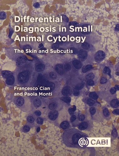
Differential Diagnosis in Small Animal Cytology. The Skin and Subcutis
Par : ,Formats :
- Paiement en ligne :
- Livraison à domicile ou en point Mondial Relay estimée à partir du 27 marsCet article sera commandé chez un fournisseur et vous sera envoyé 10 jours après la date de votre commande.
- Retrait Click and Collect en magasin gratuit
- Livraison à domicile ou en point Mondial Relay estimée à partir du 27 mars
- Réservation en ligne avec paiement en magasin :
- Indisponible pour réserver et payer en magasin
- Nombre de pages194
- FormatGrand Format
- PrésentationBroché
- Poids0.515 kg
- Dimensions18,9 cm × 24,4 cm × 1,5 cm
- ISBN978-1-78639-225-1
- EAN9781786392251
- Date de parution03/10/2019
- ÉditeurCABI
Résumé
Illustrated with high-quality photomicrographs, Differential Diagnosis in Small Animal Cytology : The Skin and Subcutis is a comprehensive resource for identifying through cytology the most common cutaneous and subcutaneous diseases of dogs and cats. With key points describing the main clinical and cytological features of each lesion, the book provides lists of differential diagnoses, including diagnostic algorithms, and handy 'pearls and pitfalls' boxes.
It is also enriched by chapters on the correct use and maintenance of the microscope, and techniques of collection and preparation of cytological specimens, making the book a valuable resource for veterinary pathologists (clinical and anatomic), residents, veterinary undergraduate students and small animal practitioners. Key features : Over 130 photomicrographs of the most common skin and subcutaneous lesions to help with diagnosis.
Ideal reference book with concise descriptions of each lesion. Organised into key bullet points to facilitate use during diagnostic work, or as a revision aid.
Vous aimerez aussi